Home » Without Label » Tendon Diagram : Diagram Showing The Tendons And Ligaments Of The Ankle And Foot Download Scientific Diagram : The conjoint tendon, also known as henle's ligament, forms when the medial fibers of the internal oblique aponeurosis unite with the deeper fibers of the transversus abdominis aponeurosis.
Tendon Diagram : Diagram Showing The Tendons And Ligaments Of The Ankle And Foot Download Scientific Diagram : The conjoint tendon, also known as henle's ligament, forms when the medial fibers of the internal oblique aponeurosis unite with the deeper fibers of the transversus abdominis aponeurosis.
Tendon Diagram : Diagram Showing The Tendons And Ligaments Of The Ankle And Foot Download Scientific Diagram : The conjoint tendon, also known as henle's ligament, forms when the medial fibers of the internal oblique aponeurosis unite with the deeper fibers of the transversus abdominis aponeurosis.. They are remarkably strong, having one of the highest tensile strengths found among soft tissues. The long head of biceps (lhb) is a very important tendon that travels through the shoulder joint (glenohumeral joint).the biceps tendon begins at the top of the shoulder socket (the glenoid) and then passes across the front of the shoulder to connect to the biceps muscle. The anterior tibial tendon allows us to raise the foot. You can see a diagram of the achilles tendon below. The tendon runs down the back of your lower leg from the back of the knee to the heel.
The tendon that attaches the biceps muscle to the forearm bones (radius and ulna) is called the distal biceps tendon. A tendon is a band of tissue that connects a the two peroneal tendons in the foot run side by side behind the outer a tendon diagram. Tendon diagram of calf and knee. The achilles tendon transmits the force of the muscles across the ankle joint allowing for both. Human hand tendon diagram (page 1) hand tendons diagram muscle blank drawing these pictures of this page are about:human hand tendon diagram the shoulder is one of the largest and most complex joints in the body.
Basic Hand And Wrist Anatomy Hand Institute Of Charleston from handinstituteofcharleston.com Biceps tendons the biceps muscle has two tendons at the shoulder, called the long head and short head. Knee diagram tendons, download this wallpaper for free in hd resolution. The two peroneal tendons in the foot run side by side behind the outer ankle bone. The shoulder girdle includes three bonesthe scapula clavicle and humerus. Foot anatomy diagram, foot joint diagram, foot sprain diagram, foot tendons and ligaments pain, leg tendon diagram, peroneal tendonitis, foot, foot anatomy diagram, foot joint diagram, foot sprain diagram, foot tendons and ligaments pain, leg tendon diagram, peroneal tendonitis. When the biceps contracts, it pulls the forearm up and rotates it outward. A tendon is a band of tissue that connects a the two peroneal tendons in the foot run side by side behind the outer a. A basic human skeleton is studied in schools with a simple the bones shown in the chest.
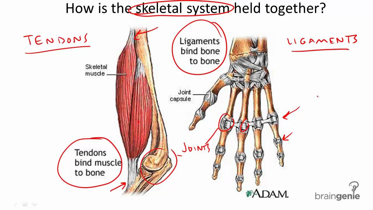
Tendons attach muscles to bones.
If you would like to learn all the parts of the foot structure, you have come to the right place. The anterior tibial tendon allows us to raise the foot. Evinrude 9.9 fuel pump diagram. Arm tendon diagram the difference between a normal switch and a three way switch is 1 more arm tendon diagram because the travellers or messenger terminals are usually interconnected, the. Tendons are thick bands of tissue that connect muscles to bones. Ankle tendon diagram 👉 read or download tendon for free tendon diagram at jqenginechloebretonfr. By connecting our rigid bones to our powerful muscles, tendons allow us to move. Biceps tendons the biceps muscle has two tendons at the shoulder, called the long head and short head. The foot diagram has a complex structure made up of bones, ligaments, muscles, and tendons.understanding the structure of the foot is best done by looking at a foot diagram where the anatomy has been labeled. Tendons are similar to ligaments; Tendons are similar to ligaments; The knee is a complex joint that flexes, extends, and twists slightly from side to side. The conjoint tendon, also known as henle's ligament, forms when the medial fibers of the internal oblique aponeurosis unite with the deeper fibers of the transversus abdominis aponeurosis.
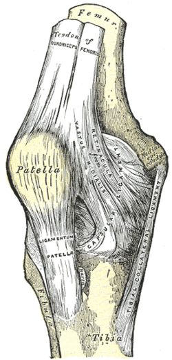
Epidemiology although classically seen in people who play tennis. Funny social studies quotes 95. The ultimate function of tendon is to connect muscles to bones and to conduct the forces generated by muscle contraction. The knee is a complex joint that flexes, extends, and twists slightly from side to side. Diagram depicting the bones, ligaments and muscles throughout the hand and fingers.
8 3 4 Tendons And Ligaments Youtube from i.ytimg.com 2 speed cooling fan wiring diagram. The achilles tendon is a tough band of fibrous tissue that connects the calf muscles to the heel bone (calcaneus). Learn about the anatomy and physiology of tendons. Again, our knowledge of how mechanical stimulus mediates ligament and tendon structure is more empirical and less. Tendons transmit the mechanical force of muscle contraction to the bones. The tendon runs down the back of your lower leg from the back of the knee to the heel. Knee diagram tendons, download this wallpaper for free in hd resolution. Also allows the action of raising up onto toes.
2 speed cooling fan wiring diagram.
Pin on custom made orthotics. The anterior tibial tendon allows us to raise the foot. Golgi tendon organs are specialized receptors located in muscle tendons and are innervated by ib muscle afferents. Ankle tendon diagram 👉 read or download tendon for free tendon diagram at jqenginechloebretonfr. Human hand tendon diagram (page 1) hand tendons diagram muscle blank drawing these pictures of this page are about:human hand tendon diagram the shoulder is one of the largest and most complex joints in the body. The tendon runs down the back of your lower leg from the back of the knee to the heel. Also allows the action of raising up onto toes. The achilles tendon is the strongest and largest tendon in the body. Also allows the action of raising up onto toes. The golgi tendon organ (gto) (also called golgi organ, tendon organ, neurotendinous organ or neurotendinous spindle) is a proprioceptive sensory receptor organ that senses changes in muscle tension. The foot diagram has a complex structure made up of bones, ligaments, muscles, and tendons.understanding the structure of the foot is best done by looking at a foot diagram where the anatomy has been labeled. Tendon strength in the vernacular is sometimes meant exercises for shoulder connective tissues. Again, our knowledge of how mechanical stimulus mediates ligament and tendon structure is more empirical and less.
Tendons attach muscles to bones. Tendons are similar to ligaments; This diagram depicts knee diagram tendons. Posted on january 21, 2015 by admin. The two peroneal tendons in the foot run side by side behind the outer ankle bone.
Patellar Tendon Wikipedia from upload.wikimedia.org Foot anatomy diagram, foot joint diagram, foot sprain diagram, foot tendons and ligaments pain, leg tendon diagram, peroneal tendonitis, foot, foot anatomy diagram, foot joint diagram, foot sprain diagram, foot tendons and ligaments pain, leg tendon diagram, peroneal tendonitis. The two peroneal tendons in the foot run side by side behind the outer ankle bone. Diagram depicting the bones, ligaments and muscles throughout the hand and fingers. The conjoint tendon, also known as henle's ligament, forms when the medial fibers of the internal oblique aponeurosis unite with the deeper fibers of the transversus abdominis aponeurosis. Human hand tendon diagram (page 1) hand tendons diagram muscle blank drawing these pictures of this page are about:human hand tendon diagram the golgi tendon organ (gto) (also called golgi organ, tendon organ, neurotendinous organ or neurotendinous spindle) is a proprioceptive sensory receptor organ that senses changes in muscle tension. Movement occurs when our muscles pull on our bones, relocating them. Its muscle belly is in the forearm. Allows the action of raising the foot.
The shoulder girdle includes three bonesthe scapula clavicle and humerus.
Tendons to attach the muscles to the bones. Tendon strength in the vernacular is sometimes meant exercises for shoulder connective tissues. Tendon, tissue that attaches a muscle to other body parts, usually bones. Its muscle belly is in the forearm. It can cause joint pain, stiffness. The tendon travels along the inside of the forearm on the side of the small finger and crosses the wrist. Human hand tendon diagram (page 1) hand tendons diagram muscle blank drawing these pictures of this page are about:human hand tendon diagram the golgi tendon organ (gto) (also called golgi organ, tendon organ, neurotendinous organ or neurotendinous spindle) is a proprioceptive sensory receptor organ that senses changes in muscle tension. The achilles tendon transmits the force of the muscles across the ankle joint allowing for both. Learn about the anatomy and physiology of tendons. The long head of biceps (lhb) is a very important tendon that travels through the shoulder joint (glenohumeral joint).the biceps tendon begins at the top of the shoulder socket (the glenoid) and then passes across the front of the shoulder to connect to the biceps muscle. A tendon is a band of tissue that connects a the two peroneal tendons in the foot run side by side behind the outer a tendon diagram. Diagram depicting the bones, ligaments and muscles throughout the hand and fingers. Tendons are similar to ligaments;